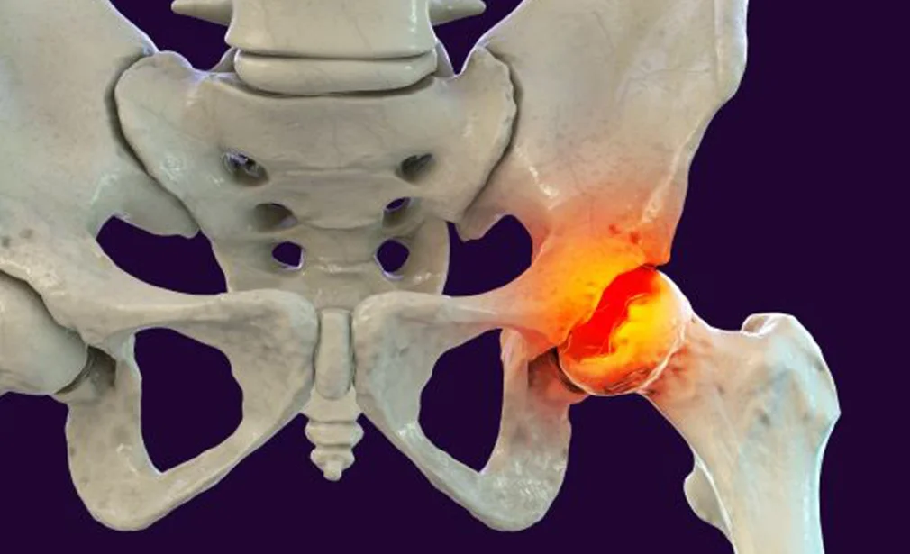
Perthes disease (Legg-Calvé-Perthes disease) is avascular necrosis of the femoral head — the blood supply to the ball of the hip joint is temporarily interrupted, causing the bone to die, collapse, and then regenerate over a period of 2–5 years.
It affects children between ages 4–10, with peak incidence at 5–7 years. Boys are affected 4–5 times more commonly than girls. It is unilateral in about 90% of cases. The disease progresses through four phases: initial necrosis → fragmentation (collapse) → re-ossification (healing) → remodelling. The goal of treatment is to maintain a spherical femoral head within a well-covered socket throughout the healing process.
The exact cause of the vascular interruption remains unknown in most cases:
- Coagulation abnormalities: Elevated levels of factor V Leiden, protein C/S deficiency, and anti-phospholipid antibodies have been found in some children — suggesting a thrombotic tendency
- Trauma: Repeated minor trauma or a single significant injury may compromise the fragile blood supply to the immature femoral head
- Delayed skeletal maturity: Children with Perthes disease often have a bone age 1–2 years behind their chronological age and are smaller than average
- Passive smoking: Associated with increased risk in some studies — suggesting a vascular mechanism
- Growth hormone deficiency: Minor association in some cases
👶
How does the child present?
- Painless limp — the most common presentation. Parents notice the child limping, often attributed to a minor fall initially
- Hip or groin pain — variable; may be felt in the knee due to referred pain along the obturator nerve
- Reduced hip range of motion — particularly internal rotation and abduction are the first movements to be lost
- Muscle spasm around the hip — the child holds the hip in external rotation
- Leg length discrepancy — from collapse of the femoral head
- Symptoms often worsen after activity and improve with rest
- Child appears generally well — no fever, normal inflammatory markers (unlike septic arthritis)
🩺
What tests are required?
- X-ray of the pelvis (AP and frog-lateral): The primary investigation — shows density changes, fragmentation, flattening, and eventually healing of the femoral head. The Catterall and Herring lateral pillar classifications grade disease severity and guide treatment
- MRI of the hip: The most sensitive early investigation — detects avascular necrosis before X-ray changes are visible. Shows the extent of necrosis and any extrusion of the femoral head
- Bone scan (technetium): reduced uptake confirms avascular necrosis when MRI is not available
- Blood tests (FBC, ESR, CRP): to exclude septic arthritis and transient synovitis — inflammatory markers are normal in Perthes disease
- Thrombophilia screening: factor V Leiden, protein C/S levels
💊
What are the treatment options?
Treatment aims to keep the femoral head contained within the acetabulum (socket) so that the healing head remodels into a spherical shape:
- Activity restriction and analgesia: Reducing high-impact activities (running, jumping) during the painful phase. Anti-inflammatory medication (ibuprofen) for pain
- Physiotherapy: Hip abduction exercises to maintain range of motion — critical throughout the disease course
- Traction and casting: For acute pain relief and to achieve abduction — Petrie casts or skin traction
- Observation (younger children with mild disease): Children under 6 with good range of motion and less than 50% lateral pillar involvement often do well with physiotherapy and monitoring alone
- Containment surgery (Herring B/C, age >6, >50% head involvement): The femoral head is redirected into the socket by:
- Femoral varus derotation osteotomy (VDRO): The femur is cut and tilted to direct the femoral head deeper into the socket
- Salter innominate osteotomy: The pelvis is redirected to provide better roof coverage over the femoral head
- Both may be combined in complex cases
🌟
What is the expected outcome?
The outcome of Perthes disease depends primarily on age at onset and severity. Children under 6 with mild-to-moderate disease have an excellent prognosis — the hip heals to a near-normal spherical shape without surgery in the majority. Older children (over 8) and those with severe head involvement (Herring C) have a higher risk of a misshapen head and early adult arthritis. Surgical containment significantly improves outcomes in this higher-risk group. Long-term follow-up until skeletal maturity is essential.