Rickets is a condition characterised by softening and weakening of the growing skeleton in children due to deficiency of Vitamin D, calcium, or phosphate — essential minerals required for proper bone mineralisation. It affects the growth plates, causing them to widen and the bones to become soft, resulting in skeletal deformity.
Rickets remains common in India, particularly in children with limited sunlight exposure, exclusively breastfed infants without supplementation, and children with poor dietary intake of Vitamin D and calcium. It is the most common cause of bowed legs in developing countries.
- Nutritional Vitamin D deficiency (most common): Inadequate sunlight exposure + insufficient dietary intake — particularly in exclusively breastfed infants, children with dark skin, and those kept indoors
- Calcium deficiency rickets: Common in India — adequate Vitamin D but insufficient dietary calcium (low dairy intake)
- Vitamin D-dependent rickets Type I (VDDR1): Genetic mutation in the enzyme converting Vitamin D to its active form (1-alpha-hydroxylase deficiency)
- Vitamin D-dependent rickets Type II (VDDR2): Resistance to active Vitamin D at the receptor level
- X-linked hypophosphataemic rickets (XLH): The most common hereditary form — phosphate-wasting through the kidney causing low phosphate levels. Does not respond to standard Vitamin D treatment
- Malabsorption: Coeliac disease, Crohn's disease, and cholestatic liver disease impair Vitamin D absorption
👶
How does the child present?
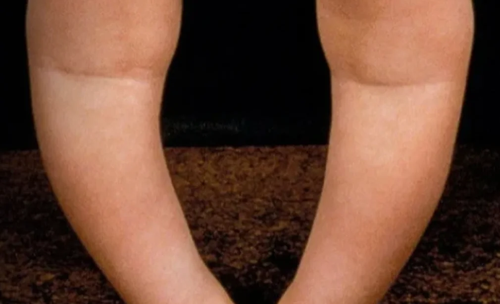
- Bowed legs (genu varum) — the hallmark in young walking children — often severe and progressive
- Knock knees (genu valgum) — can occur with calcium-deficiency rickets in older children
- Prominent wrists and ankles — "rachitic rosary" at the wrist growth plates
- Frontal bossing (prominent forehead), delayed tooth eruption, large fontanelle
- Short stature and growth failure
- Bone pain and tenderness, especially in the legs
- Muscle weakness and hypotonia — the child may appear floppy or have delayed motor milestones
- Fractures from minor trauma — pathological fractures due to bone softness
- In infants: hypocalcaemic seizures, tetany, or laryngospasm
🩺
What tests are required?
- Blood tests: Serum calcium, phosphate, alkaline phosphatase (elevated in rickets), Vitamin D (25-OH D3), PTH (parathyroid hormone), and renal function tests
- Urine tests: Urinary calcium:creatinine ratio, urinary phosphate, tubular reabsorption of phosphate (for XLH)
- X-rays: Classic findings — widening, cupping, and fraying of growth plates (especially at wrist and knee); bowing of long bones; reduced bone density; Looser zones (pseudofractures)
- Genetic testing for hereditary forms of rickets (XLH, VDDR)
- Bone age X-ray to assess skeletal maturity
- Full-length leg X-rays for deformity quantification and surgical planning
💊
What are the treatment options?
- Vitamin D and calcium supplementation: High-dose Vitamin D3 (cholecalciferol) and calcium supplementation for nutritional rickets — the mainstay of treatment. Monitored with serial blood tests. Most deformities correct spontaneously in young children once mineralisation is restored
- Phosphate supplementation + active Vitamin D (calcitriol): For X-linked hypophosphataemic rickets — oral phosphate supplements and calcitriol are required lifelong. Newer treatment with burosumab (anti-FGF23 antibody) has significantly improved outcomes
- Dietary and sunlight advice: Increasing sunlight exposure (15–30 min daily) and dietary sources of Vitamin D (fortified milk, fish, eggs) and calcium (dairy products, leafy vegetables)
- Growth modulation surgery (8-plate): For persistent angular deformity after biochemical correction in children with open growth plates — guided growth plates steer the bones to grow straight
- Corrective osteotomy: For severe persistent deformity in older children or after growth plate closure — the bone is cut and realigned
🌟
What is the expected outcome?
Nutritional rickets is entirely preventable and responds excellently to Vitamin D and calcium supplementation. In young children, skeletal deformities (bowed legs) often remodel spontaneously once the underlying deficiency is corrected, avoiding the need for surgery. Hereditary forms of rickets require long-term medical management but modern treatments including burosumab allow near-normal growth and skeletal development. Surgical correction of persistent deformity gives reliable, long-lasting results.