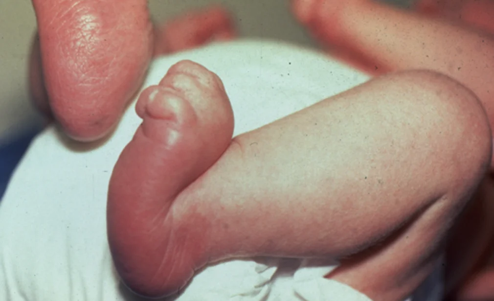
Vertical talus (also called congenital vertical talus or rocker-bottom foot) is a rare congenital foot deformity in which the talus bone is locked in a vertical position, causing the midfoot joints to dislocate. The result is a rigid, convex-soled foot that resembles the bottom of a rocking chair — hence the term "rocker-bottom foot."
Unlike flexible flat feet, vertical talus is a rigid deformity present from birth and will not resolve on its own. It requires early, structured treatment to achieve a functional, pain-free foot.
Vertical talus can be isolated or associated with underlying conditions:
- Idiopathic (isolated): Approximately half of cases have no identifiable cause and occur in otherwise healthy infants
- Neuromuscular conditions: Spina bifida, arthrogryposis, and sacral agenesis are commonly associated
- Chromosomal syndromes: Trisomy 13, 15, and 18 — genetic workup is recommended in all cases
- Intrauterine positioning: Abnormal fetal position during development can contribute
- Both feet are affected in approximately 50% of cases
👶
How does the child present?
- Characteristic "rocker-bottom" appearance of the foot — convex sole, prominent heel
- The foot is rigid — unlike flexible flat feet, the arch cannot be recreated by passive manipulation
- The forefoot is dorsiflexed (pointing upward) and the hindfoot is in equinus (pointing downward)
- Tightness of the Achilles tendon and anterior tendons
- If untreated and the child begins walking, painful calluses develop on the prominent midfoot where weight is borne abnormally
- Associated neurological or systemic features may be present depending on underlying cause
🩺
What tests are required?
- Clinical examination confirming rigidity and rocker-bottom deformity
- Lateral X-rays of the foot in forced plantar flexion — the talus remains vertical (axis greater than 45°), confirming the diagnosis
- Ultrasound can be helpful in very young infants before bones are ossified
- Genetic testing (chromosomal karyotype) to identify associated syndromes
- Neurological assessment and spine imaging (MRI) to evaluate for spinal dysraphism
- Echocardiogram and renal ultrasound as part of syndrome workup when indicated
💊
What are the treatment options?
Treatment should begin as early as possible — ideally in the first weeks of life:
- Reverse Ponseti serial casting: The foot is progressively stretched and cast in the opposite direction to club foot — casting begins from birth and continues for 4–6 weeks to stretch soft tissues and partially reduce the talonavicular joint
- Mini-open surgical reduction: After casting, a small surgical procedure is performed to fully reduce and stabilise the talonavicular joint. A temporary Kirschner wire (K-wire) holds the joint in position while healing occurs
- Achilles tenotomy: The Achilles tendon is lengthened percutaneously at the time of surgery to correct hindfoot equinus
- Post-operative casting and bracing: A full-length cast is worn for 6–8 weeks after surgery, followed by foot abduction bracing (similar to club foot protocol) to maintain correction
- Salvage procedures: For older children or failed primary treatment — more extensive surgery may be required to achieve a plantigrade foot
🌟
What is the expected outcome?
When treatment begins in early infancy using the reverse Ponseti technique followed by mini-open surgical correction, the majority of children achieve a well-aligned, plantigrade, and functional foot. Children can walk normally, wear standard footwear, and participate in age-appropriate activities. Early treatment produces significantly better outcomes than delayed intervention. Long-term follow-up is essential as the child grows to monitor for any recurrence of deformity.