Developmental Dysplasia of the Hip (DDH) is a condition where the hip joint of a newborn baby does not form correctly, leaving the ball portion of the joint loose or completely dislocated from the socket. If diagnosed early in infancy, it can be treated using simple, non-surgical braces. However, if left undiagnosed until a child starts walking, major reconstruction surgery is often required. Knowing the early warning signs is a critical skill for new parents.
What causes Developmental Dysplasia of the Hip?
The socket of a baby's hip joint is made of soft, flexible cartilage that hardens into bone over time. If the ball of the joint does not sit snugly in the socket, the socket will fail to develop its normal deep cup shape. Risk factors include breech position in the womb, family history of DDH, and tight swaddling that locks the baby's legs straight and together.
Early Warning Signs Parents Can Spot
Newborn babies are regularly screened for DDH during checkups, but parents can look for these signs at home:
- Unequal skin folds: Look at your baby's thighs and buttocks from behind. Extra or asymmetric skin creases on one side can point to a dislocated hip.
- Limited flexibility: When changing diapers, check if one leg does not spread outward as far as the other.
- Different leg lengths: One leg may appear shorter than the other.
- Clicking or popping: Feeling or hearing a click when rotating the baby's hips (though physiological clicking is common, it should always be verified by an expert).
- Limping or waddling: In older children who have started walking, a persistent limp, waddle, or walking on tiptoes on one side can indicate undiagnosed DDH.
Diagnosing DDH
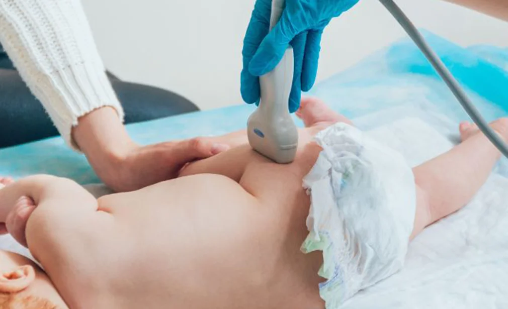
Because an infant's hips are made of cartilage, standard X-rays are not effective in babies under 4 to 6 months old. Instead, a specialized hip ultrasound (sonography) is the diagnostic test of choice. It is painless, quick, and highly accurate at demonstrating the joint alignment.
Treatments for DDH
1. The Pavlik Harness (Under 6 Months)
A soft fabric harness that keeps the baby's hips bent upward and spread outward (the "frog-leg" position). This keeps the ball pressed firmly into the socket, naturally stimulating the cartilage socket to grow into a normal, deep cup shape. It is typically worn 24 hours a day for 8 to 12 weeks.
2. Closed Reduction & Spica Casting (6 to 18 Months)
If harness treatment fails or the child is diagnosed older, a closed reduction is performed. Under anesthesia, the doctor gently manipulates the hip back into place and applies a plaster body cast (spica cast) from the chest to the ankles for 3 to 4 months to hold the joint secure.
3. Open Surgery (Over 18 Months)
For children diagnosed late, the socket has already filled with tissue. Open surgery is required to clear out the joint socket, reconstruct the bones, and secure the hip, followed by spica casting.
