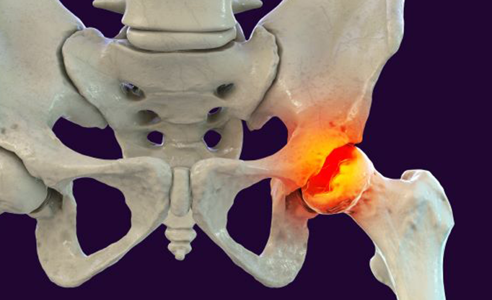
What it means?
It is a condition characterized structural damage to the ball of the hip joint (femoral head) in the childhood secondary to lack of blood flow to the femoral head.
What causes it?
The exact cause for this condition is not known. Blood flow to the ball of the hip joint (femoral head) is compromised. As a result of this, bone cells in the femoral head dies due to lack of blood supply. The dead cells remain inside the femoral head which makes that region soft. This soft portion of the femoral head cannot bear the load of body weight which gets deformed on bearing weight over the affected hip joint. This deformed ball of the hip joint will cause pain and restriction of the hip joint movements. Over a period of time, gradually the blood flow will get restored and dead bone will be replaced by new bone and ball of the hip joint will return to normal status if not got deformed in the previous stages.
How child will present like?
- More common in boys
- Usually occurs in child aged between 4 to 10 years
- Child present with limp on the affected side while walking
- Child will complain of pain in the hip on activities also pain radiating from the hip to the inner side of the knee joint
- Stiffness of the affected hip joint
What are the tests required?
- Child need to be checked clinically for his/ her hip range of movements, any discomfort in the hip joint, limb length difference, walking pattern
- X-ray of both the hip joint helps to assess the status of femoral head, child is in which stage of the disease, to plan the further management. Serial x-ray also required to assess the progression of the disease.
What are the treatment options?
- Medications like analgesics helps to relieve the pain symptoms
- Complete bed rest and skin traction to provide rest to the affected joint and relieve muscle spasm and child advised not to bear weight over the affected limb
- Physical therapy helps to maintain the hip range of movements
- In early stage, child can be taken under anaesthesia, hip joint range of movement and proper positioning of femoral head in the hip joint were assessed after injecting the dye into the hip joint. This also provides information regarding the shape of the head. If the groin muscles were found to be tight and restricting the ball from going into hip joint, tight muscle can be released and get the hip into better position and maintained in the same position with the help of abduction cast for 6 weeks then hip range of movement will be started
- In more severe cases, the child may require surgery to keep the ball into the socket of the hip joint which can be achieved by cutting the thigh bone(femur) or the pelvis or both. After achieving correction the bones were fixed with metal plate and screws to hold everything in place
What will be outcome?
- Most of the children diagnosed early and treated according to the stage of the disease will do well and will return to normal activity
- Younger children and in hips have all range of movement will have better outcome
- Child whose ball of the hip joint (femoral head) is not round can develop pain and arthritis of the hip joint later age which may require further treatment in the future